Intent: The items in this section document the risk, presence, appearance, and change of pressure ulcers/injuries. This section also notes other skin ulcers, wounds, or lesions, and documents some treatment categories related to skin injury or avoiding injury. It is important to recognize and evaluate each resident’s risk factors and to identify and evaluate all areas at risk of constant pressure. A complete assessment of skin is essential to an effective pressure ulcer prevention and skin treatment program. Be certain to include in the assessment process, a holistic approach. It is imperative to determine the etiology of all wounds and lesions, as this will determine and direct the proper treatment and management of the wound.
CMS is aware of the array of terms used to describe alterations in skin integrity due to pressure. Some of these terms include: pressure ulcer, pressure injury, pressure sore, decubitus ulcer, and bed sore. Acknowledging that clinicians may use and documentation may reflect any of these terms, it is acceptable to code pressure-related skin conditions in Section M if different terminology is recorded in the clinical record, as long as the primary cause of the skin alteration is related to pressure. For example, if the medical record reflects the presence of a Stage 2 pressure injury, it should be coded on the MDS as a Stage 2 pressure ulcer.
M0100: Determination of Pressure Ulcer/Injury Risk
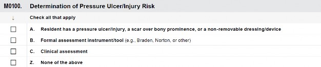
Pressure ulcers/injuries occur when tissue is compressed between a bony prominence and an external surface. In addition to pressure, shear force, and friction are important contributors to pressure ulcer/injury development.
The underlying health of a resident’s soft tissue affects how much pressure, shear force, or friction is needed to damage tissue. Skin and soft tissue changes associated with aging, illness, small blood vessel disease, and malnutrition increase vulnerability to pressure ulcers/injuries.
Additional external factors, such as excess moisture, microclimate, and tissue exposure to urine or feces, can increase risk.
The care planning process should include efforts to stabilize, reduce, or remove underlying risk factors; to monitor the impact of the interventions; and to modify the interventions as appropriate based on the individualized needs of the resident.
M0100: Determination of Pressure Ulcer/Injury Risk (cont.)
DEFINITION
HEALED PRESSURE ULCER
Completely closed, fully epithelialized, covered completely with epithelial tissue, or resurfaced with new skin, even if the area continues to have some surface discoloration.
DEFINITION
HEALED PRESSURE ULCER
Completely closed, fully epithelialized, covered completely with epithelial tissue, or resurfaced with new skin, even if the area continues to have some surface discoloration.
Throughout this section, terminology referring to “healed” versus “unhealed” ulcers refers to whether or not the ulcer is “closed” versus “open.” When considering this, recognize that Stage 1, Deep Tissue Injury (DTI), and unstageable pressure ulcers although “closed” (i.e., may be covered with tissue, eschar, slough, etc.) would not be considered “healed.”
Facilities should be aware that the resident is at higher risk of having the area of a closed pressure ulcer open up due to damage, injury, or pressure, because of the loss of tensile strength of the overlying tissue. Tensile
strength of the skin overlying a closed pressure ulcer is 80% of normal skin tensile strength. Facilities should put preventative measures in place that will mitigate the opening of a closed ulcer due to the fragility of the overlying tissue.
DEFINITIONS
PRESSURE ULCER/ INJURY RISK FACTOR
Examples of risk factors include immobility and decreased functional ability; co-morbid conditions such as end-stage renal disease, thyroid disease, or diabetes; drugs such as steroids; impaired diffuse or localized blood flow; resident refusal of care and treatment; cognitive impairment; exposure of skin to urinary and fecal incontinence; microclimate, malnutrition, and hydration deficits; and a healed ulcer.
PRESSURE ULCER/ INJURY RISK TOOLS
Screening tools that are designed to help identify residents who might develop a pressure ulcer/injury. A common risk assessment tool is the Braden Scale for Predicting Pressure Sore Risk©.
DEFINITIONS
PRESSURE ULCER/ INJURY RISK FACTOR
Examples of risk factors include immobility and decreased functional ability; co-morbid conditions such as end-stage renal disease, thyroid disease, or diabetes; drugs such as steroids; impaired diffuse or localized blood flow; resident refusal of care and treatment; cognitive impairment; exposure of skin to urinary and fecal incontinence; microclimate, malnutrition, and hydration deficits; and a healed ulcer.
PRESSURE ULCER/ INJURY RISK TOOLS
Screening tools that are designed to help identify residents who might develop a pressure ulcer/injury. A common risk assessment tool is the Braden Scale for Predicting Pressure Sore Risk©.
Review the medical record, including skin care flow sheets or other skin tracking forms, nurses’ notes, and pressure ulcer/injury risk assessments.
Speak with the treatment nurse and direct care staff on all shifts to confirm conclusions from the medical record review and observations of the resident.
Examine the resident and determine whether any ulcers, injuries, scars, or non-removable dressings/devices are present. Assess key areas for pressure ulcer/injury development (e.g., sacrum, coccyx, trochanters, ischial tuberosities, and heels). Also assess bony prominences (e.g., elbows and ankles) and skin that is under braces or subjected to pressure (e.g., ears from oxygen tubing).
Coding Instructions
For this item, check all that apply:
Check A if resident has a Stage 1 or greater pressure ulcer/injury, a scar over bony prominence, or a non-removable dressing/ device. Review descriptions of pressure ulcers/injuries and information obtained during physical examination and medical record review. Examples of non-removable dressings/devices include a primary surgical dressing, a cast, or a brace.
M0100: Determination of Pressure Ulcer/Injury Risk (cont.)
Check B if a formal assessment has been completed. An example of an established pressure ulcer risk tool is the Braden Scale for Predicting Pressure Sore Risk©. Other tools may be used.
Check C if the resident’s risk for pressure ulcer/injury development is based on clinical assessment. A clinical assessment could include a head-to-toe physical examination of the skin and observation or medical record review of pressure ulcer/injury risk factors. Examples of risk factors include the following:
impaired/decreased mobility and decreased functional ability
co-morbid conditions, such as end stage renal disease, thyroid disease, or diabetes mellitus;
drugs, such as steroids, that may affect wound healing;
impaired diffuse or localized blood flow (e.g., generalized atherosclerosis or lower extremity arterial insufficiency);
resident refusal of some aspects of care and treatment;
cognitive impairment;
urinary and fecal incontinence;
malnutrition and hydration deficits; and
healed pressure ulcers, especially Stage 3 or 4 which are more likely to have recurrent breakdown.
Check Z if none of the above apply.
M0150: Risk of Pressure Ulcers/Injuries

It is important to recognize and evaluate each resident’s risk factors and to identify and evaluate all areas at risk of constant pressure.
The care process should include efforts to stabilize, reduce, or remove underlying risk factors; to monitor the impact of the interventions; and to modify the interventions as appropriate.
Based on the item(s) reviewed for M0100, determine if the resident is at risk for developing a pressure ulcer/injury.
M0150: Risk of Pressure Ulcers/Injuries (cont.)
If the medical record reveals that the resident currently has a pressure ulcer/injury, a scar over a bony prominence, or a non-removable dressing or device, the resident is at risk for worsening or new pressure ulcers/injuries.
Review formal risk assessment tools to determine the resident’s “risk score.”
Review the components of the clinical assessment conducted for evidence of pressure ulcer/injury risk.
Coding Instructions
Code 0, no: if the resident is not at risk for developing pressure ulcers/injuries based on a review of information gathered for M0100.
Code 1, yes: if the resident is at risk for developing pressure ulcers/injuries based on a review of information gathered for M0100.
M0210: Unhealed Pressure Ulcers/Injuries

DEFINITION
PRESSURE ULCER/INJURY
A pressure ulcer/injury is localized injury to the skin and/or underlying tissue, usually over a bony prominence, as a result of intense and/or prolonged pressure or pressure in combination with shear. The pressure ulcer/injury can present as intact skin or an open ulcer and may be painful.
DEFINITION
PRESSURE ULCER/INJURY
A pressure ulcer/injury is localized injury to the skin and/or underlying tissue, usually over a bony prominence, as a result of intense and/or prolonged pressure or pressure in combination with shear. The pressure ulcer/injury can present as intact skin or an open ulcer and may be painful.
Pressure ulcers/injuries and other wounds or lesions affect quality of life for residents because they may limit activity, may be painful, and may require time- consuming treatments and dressing changes.
The pressure ulcer/injury definitions used in the RAI Manual have been adapted from those recommended by the National Pressure Ulcer Advisory Panel (NPUAP) 2016 Pressure Injury Staging System.
An existing pressure ulcer/injury identifies residents at risk for further complications or skin injury. Risk factors described in M0100 should be addressed.
For MDS assessment, initial numerical staging of
pressure ulcers and the initial numerical staging of ulcers after debridement, or DTI that declares itself, should be coded in terms of what is assessed (seen or palpated, i.e. visible tissue, palpable bone) during the look-back period. Nursing homes may adopt the NPUAP guidelines in their clinical practice and nursing documentation. However, since CMS has adapted the NPUAP guidelines for MDS purposes, the definitions do not perfectly correlate with each stage as described by NPUAP. Therefore, you must code the MDS according to the instructions in this manual.
M0210: Unhealed Pressure Ulcers/Injuries (cont.)
Pressure ulcer/injury staging is an assessment system that provides a description and classification based on visual appearance and/or anatomic depth of soft tissue damage. This tissue damage can be visible or palpable in the ulcer bed. Pressure ulcer/injury staging also informs expectations for healing times.
The comprehensive care plan should be reevaluated to ensure that appropriate preventative measures and pressure ulcer/injury management principles are being adhered to when new pressure ulcers/injuries develop or when existing pressure ulcers/injuries worsen.
Review the medical record, including skin care flow sheets or other skin tracking forms.
Speak with direct care staff and the treatment nurse to confirm conclusions from the medical record review.
Examine the resident and determine whether any skin ulcers/injuries are present.
Key areas for pressure ulcer/injury development include the sacrum, coccyx, trochanters, ischial tuberosities, and heels. Other areas, such as bony deformities, skin under braces, and skin subjected to excess pressure, shear, or friction, are also at risk for pressure ulcers/injuries.
Without a full body skin assessment, a pressure ulcer/injury can be missed.
Examine the resident in a well-lit room. Adequate lighting is important for detecting skin changes. For any pressure ulcers/injuries identified, measure and record the deepest anatomical stage.
Identify any known or likely unstageable pressure ulcers/injuries.
Code based on the presence of any pressure ulcer/injury (regardless of stage) in the past 7 days.
Code 0, no: if the resident did not have a pressure ulcer/injury in the 7-day look-back period. Then skip to M1030, Number of Venous and Arterial Ulcers.
Code 1, yes: if the resident had any pressure ulcer/injury (Stage 1, 2, 3, 4, or unstageable) in the 7-day look-back period. Proceed to M0300, Current Number of Unhealed Pressure Ulcers/Injuries at Each Stage.
If an ulcer/injury arises from a combination of factors that are primarily caused by pressure, then the area should be included in this section as a pressure ulcer/injury.
Mucosal ulcers caused by pressure should not be coded in Section M. Oral mucosal ulcers are captured in item L0200C, Abnormal mouth tissue.
Mucosal pressure ulcers are not staged using the skin pressure ulcer staging system because anatomical tissue comparisons cannot be made. Therefore, mucosal ulcers (for example, those related to nasogastric tubes, nasal oxygen tubing, endotracheal tubes, urinary catheters, etc.) should not be coded here.
M0210: Unhealed Pressure Ulcers/Injuries (cont.)
If a pressure ulcer is surgically closed with a flap or graft, it should be coded as a surgical wound and not as a pressure ulcer. If the flap or graft fails, continue to code it as a surgical wound until healed.
Residents with diabetes mellitus (DM) can have a pressure, venous, arterial, or diabetic neuropathic ulcer. The primary etiology should be considered when coding whether a resident with DM has an ulcer/injury that is caused by pressure or other factors.
If a resident with DM has a heel ulcer/injury from pressure and the ulcer/injury is present in the 7-day look-back period, code 1 and proceed to code items in M0300 as appropriate for the pressure ulcer/injury.
If a resident with DM has an ulcer on the plantar (bottom) surface of the foot closer to the metatarsals and the ulcer is present in the 7-day look-back period, code 0 and proceed to M1040 to code the ulcer as a diabetic foot ulcer. It is not likely that pressure is the primary cause of the resident’s ulcer when the ulcer is in this location.
Scabs and eschar are different both physically and chemically. Eschar is a collection of dead tissue within the wound that is flush with the surface of the wound. A scab is made up of dried blood cells and serum, sits on the top of the skin, and forms over exposed wounds such as wounds with granulating surfaces (like pressure ulcers, lacerations, evulsions, etc.). A scab is evidence of wound healing. A pressure ulcer that was staged as a 2 and now has a scab indicates it is a healing stage 2, and therefore, staging should not change. Eschar characteristics and the level of damage it causes to tissues is what makes it easy to distinguish from a scab. It is extremely important to have staff who are trained in wound assessment and who are able to distinguish scabs from eschar.
If two pressure ulcers/injuries occur on the same bony prominence and are separated, at least superficially, by skin, then count them as two separate pressure ulcers/injuries. Stage and measure each pressure ulcer/injury separately.
If a resident had a pressure ulcer/injury that healed during the look-back period of the current assessment, do not code the ulcer/injury on the assessment.
Skin changes at the end of life (SCALE), also referred to as Kennedy Terminal Ulcers (KTUs) and skin failure, are not primarily caused by pressure and are not coded in Section M.
M0300: Current Number of Unhealed Pressure Ulcers/Injuries at Each Stage
Steps for completing M0300A–G
For each pressure ulcer, determine the deepest anatomical stage. At admission, code based on findings from the first skin assessment that is conducted on or after and as close to the admission as possible. Do not reverse or back stage. Consider current and historical levels of tissue involvement.
For each pressure ulcer, determine the deepest anatomical stage. At admission, code based on findings from the first skin assessment that is conducted on or after and as close to the admission as possible. Do not reverse or back stage. Consider current and historical levels of tissue involvement.
Observe and palpate the base of any identified pressure ulcers present to determine the anatomic depth of soft tissue damage involved.
Ulcer staging should be based on the ulcer’s deepest anatomic soft tissue damage that is visible or palpable. If a pressure ulcer’s tissues are obscured such that the depth of soft tissue damage cannot be observed, it is considered to be unstageable (see Step 2 below).
DEFINITIONS
EPITHELIAL TISSUE
New skin that is light pink and shiny (even in persons with darkly pigmented skin). In Stage 2 pressure ulcers, epithelial tissue is seen in the center and at the edges of the ulcer. In full thickness Stage 3 and 4 pressure ulcers, epithelial tissue advances from the edges of the wound.
GRANULATION TISSUE
Red tissue with “cobblestone” or bumpy appearance; bleeds easily when injured.
DEFINITIONS
EPITHELIAL TISSUE
New skin that is light pink and shiny (even in persons with darkly pigmented skin). In Stage 2 pressure ulcers, epithelial tissue is seen in the center and at the edges of the ulcer. In full thickness Stage 3 and 4 pressure ulcers, epithelial tissue advances from the edges of the wound.
GRANULATION TISSUE
Red tissue with “cobblestone” or bumpy appearance; bleeds easily when injured.
Review the history of each pressure ulcer in the medical record. If the stageable pressure ulcer has ever been classified at a higher numerical stage than what is observed now, it should continue to be classified at the higher numerical stage until healed unless it becomes unstageable. Nursing homes that carefully document and track pressure ulcers will be able to more accurately code this item.
Pressure ulcers do not heal in a reverse sequence, that is, the body does not replace the types and layers of tissue (e.g., muscle, fat, and dermis) that were lost during pressure ulcer development before they re-epithelialize. Stage 3 and 4 pressure ulcers fill with granulation tissue. This replacement tissue is never as strong as the tissue that was lost and hence is more prone to future breakdown.
Clinical standards do not support reverse staging or back- staging as a way to document healing, as it does not accurately characterize what is occurring physiologically as the ulcer heals. For example, over time, even though a Stage 4 pressure ulcer has been healing and contracting such that it is less deep, wide, and long, the tissues that were lost (muscle, fat, dermis) will never be replaced with the same type of tissue. Previous standards using reverse staging or back-staging would have permitted identification of such a pressure ulcer as a Stage 3, then a Stage 2, and so on, when it reached a depth consistent with these stages.
Clinical standards now would require that this ulcer
continue to be documented as a Stage 4 pressure ulcer until it has completely healed unless it becomes unstageable. Nursing homes can document the healing of pressure ulcers using descriptive characteristics of the wound (i.e., depth, width, presence or absence of granulation tissue, etc.) or by using a validated pressure ulcer healing tool.
M0300: Current Number of Unhealed Pressure Ulcers/Injuries at Each Stage (cont.)
Once a pressure ulcer has healed, it is documented as a healed pressure ulcer at its highest numerical stage—in this example, a healed Stage 4 pressure ulcer. For care planning purposes, this healed Stage 4 pressure ulcer would remain at increased risk for future breakdown or injury and would require continued monitoring and preventative care.
A previously closed pressure ulcer that opens again should be reported at its worst stage, unless currently presenting at a higher stage or unstageable.
Visualization of the wound bed is necessary for accurate staging.
If, after careful cleansing of the pressure ulcer/injury, a pressure ulcer’s/injury’s anatomical tissues are obscured such that the extent of soft tissue damage cannot be observed or palpated, the pressure ulcer/injury is considered unstageable.
Pressure ulcers that have eschar (tan, black, or brown) or slough (yellow, tan, gray, green or brown) tissue present such that the anatomic depth of soft tissue damage cannot be visualized or palpated in the wound bed, should be classified as unstageable, as illustrated at https://cdn.ymaws.com/npiap.com/resource/resmgr/NPIAP-Staging-Poster.pdf.
If the wound bed is only partially covered by eschar or slough, and the anatomical depth of tissue damage can be visualized or palpated, numerically stage the ulcer, and do not code this as unstageable.
A pressure injury with intact skin that is a deep tissue injury (DTI) should not be coded as a Stage 1 pressure injury. It should be coded as unstageable, as illustrated at https://cdn.ymaws.com/npiap.com/resource/resmgr/NPIAP-Staging-Poster.pdf.
Known pressure ulcers/injuries covered by a non-removable dressing/device (e.g., primary surgical dressing, cast) should be coded as unstageable. “Known” refers to when documentation is available that says a pressure ulcer/injury exists under the non-removable dressing/device.
Step 3: Determine “Present on Admission”
For each pressure ulcer/injury, determine if the pressure ulcer/injury was present at the time of admission/entry or reentry and not acquired while the resident was in the care of the nursing home. Consider current and historical levels of tissue involvement.
DEFINITION
ON ADMISSION
As close to the actual time of admission as possible.
For each pressure ulcer/injury, determine if the pressure ulcer/injury was present at the time of admission/entry or reentry and not acquired while the resident was in the care of the nursing home. Consider current and historical levels of tissue involvement.
DEFINITION
ON ADMISSION
As close to the actual time of admission as possible.
Review the medical record for the history of the ulcer/injury.
Review for location and stage at the time of admission/entry or reentry.
If the pressure ulcer/injury was present on admission/entry or reentry and subsequently increased in numerical stage during the resident’s stay, the pressure ulcer is coded at that higher stage, and that higher stage should not be considered as “present on admission.”
If a numerically stageable pressure ulcer/injury was present on admission/entry or reentry and becomes unstageable due to slough or eschar, during the resident’s stay, the pressure ulcer/injury is coded at M0300F and should not be coded as “present on admission.”
M0300: Current Number of Unhealed Pressure Ulcers/Injuries at Each Stage (cont.)
If the pressure ulcer/injury was unstageable on admission/entry or reentry, then becomes numerically stageable later, it should be considered as “present on admission” at the stage at which it first becomes numerically stageable. If it subsequently increases in numerical stage, that higher stage should not be coded as “present on admission.”
If a resident who has a pressure ulcer/injury that was originally acquired in the facility is hospitalized and returns with that pressure ulcer/injury at the same numerical stage, the pressure ulcer/injury should not be coded as “present on admission” because it was present and acquired at the facility prior to the hospitalization.
If a resident who has a pressure ulcer/injury that was “present on admission” (not acquired in the facility) is hospitalized and returns with that pressure ulcer/injury at the same numerical stage, the pressure ulcer is still coded as “present on admission” because it was originally acquired outside the facility and has not changed in stage.
If a resident who has a pressure ulcer/injury is hospitalized and the ulcer/injury increases in numerical stage or becomes unstageable due to slough or eschar during the hospitalization, it should be coded as “present on admission” upon reentry.
If a pressure ulcer was numerically staged, then became unstageable, and is subsequently debrided sufficiently to be numerically staged, compare its numerical stage before and after it was unstageable. If the numerical stage has increased, code this pressure ulcer as not “present on admission.”
If a resident has a pressure ulcer/injury that was documented on admission then closed that reopens at the same stage (i.e., not a higher stage), the ulcer/injury is coded as “present on admission.”
If two pressure ulcers merge, that were both “present on admission,” continue to code the merged pressure ulcer as “present on admission.” Although two merged pressure ulcers might increase the overall surface area of the ulcer, there needs to be an increase in numerical stage or a change to unstageable due to slough or eschar in order for it to be considered not “present on admission.”
If a pressure ulcer/injury was unstageable on admission/entry or reentry and then becomes unstageable for another reason, it should be considered “present on admission” at the new unstageable status. For example, if a resident is admitted with a deep tissue injury, but later the injury opens, the wound bed is covered with slough, and the wound is still unstageable, this wound would still be considered “present on admission.”
M0300: Current Number of Unhealed Pressure Ulcers/Injuries at Each Stage (cont.)
Resident K is admitted to the facility without a pressure ulcer/injury. During the stay, they develop a stage 2 pressure ulcer. This is a facility acquired pressure ulcer and was not “present on admission.” Resident K is hospitalized and returns to the facility with the same stage 2 pressure ulcer. This pressure ulcer was originally acquired in the nursing home and should not be considered as “present on admission” when they return from the hospital.

Resident J is a new admission to the facility and is admitted with a stage 2 pressure ulcer. This pressure ulcer is considered as “present on admission” as it was not acquired in the facility. Resident J is hospitalized and returns with the same stage 2 pressure ulcer, unchanged from the prior admission/entry. This pressure ulcer is still considered “present on admission” because it was originally acquired outside the facility and has not changed.

M0300A: Number of Stage 1 Pressure Injuries

DEFINITIONS
STAGE 1 PRESSURE INJURY
An observable, pressure- related alteration of intact skin whose indicators, as compared to an adjacent or opposite area on the body, may include changes in one or more of the following parameters: skin temperature (warmth or coolness); tissue consistency (firm or boggy); sensation (pain, itching); and/or a defined area of persistent redness in lightly pigmented skin, whereas in darker skin tones, the injury may appear with persistent red, blue, or purple hues.
NON-BLANCHABLE
Reddened areas of tissue that do not turn white or pale when pressed firmly with a finger or device.
DEFINITIONS
STAGE 1 PRESSURE INJURY
An observable, pressure- related alteration of intact skin whose indicators, as compared to an adjacent or opposite area on the body, may include changes in one or more of the following parameters: skin temperature (warmth or coolness); tissue consistency (firm or boggy); sensation (pain, itching); and/or a defined area of persistent redness in lightly pigmented skin, whereas in darker skin tones, the injury may appear with persistent red, blue, or purple hues.
NON-BLANCHABLE
Reddened areas of tissue that do not turn white or pale when pressed firmly with a finger or device.
Stage 1 pressure injuries may deteriorate to more severe pressure ulcers/injuries without adequate intervention; as such, they are an important risk factor for further tissue damage.
Development of a Stage 1 pressure injury should be one of multiple factors that initiate pressure ulcer/injury prevention interventions.
Perform head-to-toe assessment. Conduct a full body skin assessment focusing on bony prominences and pressure- bearing areas (sacrum, buttocks, heels, ankles, etc.).
For the purposes of coding, determine that the lesion being assessed is primarily related to pressure and that other conditions have been ruled out. If pressure is not the primary cause, do not code here.
Reliance on only one descriptor is inadequate to determine the staging of a pressure injury between Stage 1 and deep tissue injury (see definition of “deep tissue injury” on page M-24). The descriptors are similar for these two types of injuries (e.g., temperature [warmth or coolness]; tissue consistency [firm or boggy]).
Check any reddened areas for ability to blanch by firmly pressing a finger into the reddened tissues and then removing it. In non-blanchable reddened areas, there is no loss of skin color or pressure-induced pallor at the compressed site.
Search for other areas of skin that differ from surrounding tissue that may be painful, firm, soft, warmer, or cooler compared to adjacent tissue. Stage 1 may be difficult to detect in individuals with dark skin tones. Visible blanching may not be readily apparent in darker skin tones. Look for temperature or color changes as well as surrounding tissue that may be painful, firm, or soft.
M0300A: Number of Stage 1 Pressure Injuries (cont.)
Enter the number of Stage 1 pressure injuries that are currently present.
Enter 0 if no Stage 1 pressure injuries are currently present.
M0300B: Stage 2 Pressure Ulcers

DEFINITION
STAGE 2 PRESSURE ULCER
Partial thickness loss of dermis presenting as a shallow open ulcer with a red-pink wound bed, without slough or bruising.
May also present as an intact or open/ ruptured blister.
DEFINITION
STAGE 2 PRESSURE ULCER
Partial thickness loss of dermis presenting as a shallow open ulcer with a red-pink wound bed, without slough or bruising.
May also present as an intact or open/ ruptured blister.
Stage 2 pressure ulcers may worsen without proper interventions.
These residents are at risk for further complications or skin injury.
Most Stage 2 pressure ulcers should heal in a reasonable time frame (e.g., 60 days).
If a pressure ulcer fails to show some evidence toward healing within 14 days, the pressure ulcer (including potential complications) and the patient’s overall clinical condition should be reassessed.
Stage 2 pressure ulcers are often related to friction and/or shearing force, and the care plan should incorporate efforts to limit these forces on the skin and tissues.
Stage 2 pressure ulcers may be more likely to heal with treatment than higher stage pressure ulcers.
The care plan should include individualized interventions and evidence that the interventions have been monitored and modified as appropriate.
M0300B: Stage 2 Pressure Ulcers (cont.)
Perform head-to-toe assessment. Conduct a full body skin assessment focusing on bony prominences and pressure-bearing areas (sacrum, buttocks, heels, ankles, etc.).
For the purposes of coding, determine that the lesion being assessed is primarily related to pressure and that other conditions have been ruled out. If pressure is not the primary cause, do not code here.
Examine the area adjacent to or surrounding an intact blister for evidence of tissue damage. If other conditions are ruled out and the tissue adjacent to or surrounding the blister demonstrates signs of tissue damage (e.g., color change, tenderness, bogginess or firmness, warmth or coolness), these characteristics suggest a deep tissue injury (DTI) rather than a Stage 2 pressure ulcer.
Stage 2 pressure ulcers will generally lack the surrounding characteristics found with a deep tissue injury.
Identify the number of these pressure ulcers that were present on admission/entry or reentry (see instructions on page M-8).
Enter the number of pressure ulcers that are currently present and whose deepest anatomical stage is Stage 2.
Enter 0 if no Stage 2 pressure ulcers are present and skip to M0300C, Stage 3.
Enter the number of these Stage 2 pressure ulcers that were first noted at the time of admission/entry AND—for residents who are reentering the facility after a hospital stay, enter the number of Stage 2 pressure ulcers that were acquired during the hospitalization (i.e., the Stage 2 pressure ulcer was not acquired in the nursing facility prior to admission to the hospital).
Enter 0 if no Stage 2 pressure ulcers were first noted at the time of admission/entry or reentry.
Stage 2 pressure ulcers by definition have partial thickness loss of the dermis. Granulation tissue, slough, and eschar are not present in Stage 2 pressure ulcers.
Do not code skin tears, tape burns, moisture associated skin damage, or excoriation here.
When a pressure ulcer presents as an intact blister, examine the adjacent and surrounding area for signs of deep tissue injury. When a deep tissue injury is determined, do not code as a Stage 2.
M0300C: Stage 3 Pressure Ulcers

DEFINITION
STAGE 3 PRESSURE ULCER
Full thickness tissue loss. Subcutaneous fat may be visible but bone, tendon or muscle is not exposed.
Slough may be present but does not obscure the depth of tissue loss. May include undermining or tunneling (see definition of undermining and tunneling on page M-20).
DEFINITION
STAGE 3 PRESSURE ULCER
Full thickness tissue loss. Subcutaneous fat may be visible but bone, tendon or muscle is not exposed.
Slough may be present but does not obscure the depth of tissue loss. May include undermining or tunneling (see definition of undermining and tunneling on page M-20).
Pressure ulcers affect quality of life for residents because they may limit activity, may be painful, and may require time-consuming treatments and dressing changes.
Pressure ulcers at more advanced stages typically require more aggressive interventions, including more frequent repositioning, attention to nutritional status, and care that may be more time or staff intensive.
An existing pressure ulcer may put residents at risk for further complications or skin injury.
If a pressure ulcer fails to show some evidence toward healing within 14 days, the pressure ulcer (including potential complications) and the resident’s overall clinical condition should be reassessed.
Tissue characteristics of pressure ulcers should be considered when determining treatment options and choices.
Changes in tissue characteristics over time are indicative of wound healing or degeneration.
Perform head-to-toe assessment. Conduct a full body skin assessment focusing on bony prominences and pressure-bearing areas (sacrum, buttocks, heels, ankles, etc.).
For the purposes of coding, determine that the lesion being assessed is primarily related to pressure and that other conditions have been ruled out. If pressure is not the primary cause, do not code here.
Identify all Stage 3 pressure ulcers currently present.
Identify the number of these pressure ulcers that were present on admission/entry or reentry.
M0300C: Stage 3 Pressure Ulcers (cont.)
Enter the number of pressure ulcers that are currently present and whose deepest anatomical stage is Stage 3.
Enter 0 if no Stage 3 pressure ulcers are present and skip to M0300D, Stage 4.
Enter the number of these Stage 3 pressure ulcers that were first noted at Stage 3 at the time of admission/entry AND—for residents who are reentering the facility after a hospital stay, enter the number of Stage 3 pressure ulcers that were acquired during the hospitalization (i.e., the Stage 3 pressure ulcer was not acquired in the nursing facility prior to admission to the hospital).
Enter 0 if no Stage 3 pressure ulcers were first noted at the time of admission/entry or reentry.
The depth of a Stage 3 pressure ulcer varies by anatomical location. Stage 3 pressure ulcers can be shallow, particularly on areas that do not have subcutaneous tissue, such as the bridge of the nose, ear, occiput, and malleolus.
In contrast, areas of significant adiposity can develop extremely deep Stage 3 pressure ulcers. Therefore, observation and assessment of skin folds should be part of overall skin assessment. Do not code moisture-associated skin damage or excoriation here.
Bone/tendon/muscle is not visible or directly palpable in a Stage 3 pressure ulcer.
M0300C: Stage 3 Pressure Ulcers (cont.)
A pressure ulcer described as a Stage 2 was noted and documented in the resident’s medical record on admission. On a later assessment, the wound is noted to be a full thickness ulcer without exposed bone, tendon, or muscle, thus it is now a Stage 3 pressure ulcer in the same location.
Coding: The admission coding would be M0300B1 as 1, and M0300B2 as 1, present upon admission/entry or reentry. On the current assessment, the coding for the Stage 2 data elements would be M0300B1 as 0, and M0300B2 is skipped, since there is no longer a Stage 2 pressure ulcer. The Stage 3 pressure ulcer currently assessed would be coded at M0300C1 as 1, and at M0300C2 as 0, not present on admission/entry or reentry.
Rationale: The designation of “present on admission” requires that the pressure ulcer be at the same location and not have increased in numerical stage or become unstageable due to slough or eschar. This pressure ulcer worsened from Stage 2 to Stage 3 after admission. M0300C1 is coded as 1 and M0300C2 is coded as 0 on the current assessment because the ulcer was not a Stage 3 pressure ulcer on admission.
A resident develops a Stage 2 pressure ulcer while at the nursing facility. The resident is discharged to an acute-care hospital and was hospitalized. The resident returns to the nursing facility with a Stage 3 pressure ulcer in the same location.
Coding: The Stage 3 pressure ulcer, assessed on reentry, would be coded at M0300C1 as 1, and at M0300C2 as 1, present on admission/entry or reentry.
Rationale: The resident developed a Stage 2 pressure ulcer while at the nursing facility. This is a “facility acquired” pressure ulcer and was not “present on admission.” The resident is hospitalized and returns with a pressure ulcer in the same location, which has now worsened to a Stage 3. Although the pressure ulcer was originally acquired in the nursing facility, it is coded as “present on admission/entry or reentry,” because it increased in numerical stage while the resident was in the hospital.
M0300C: Stage 3 Pressure Ulcers (cont.)
On admission, the resident has three small Stage 2 pressure ulcers on their coccyx. Two weeks later, the coccyx is assessed. Two of the Stage 2 pressure ulcers have merged and the third has increased in numerical stage to a Stage 3 pressure ulcer.
Coding: The admission coding would be M0300B1 as 3, and M0300B2 as 1, present on admission/entry or reentry. On the subsequent assessment, the two merged pressure ulcers would be coded at M0300B1 as 1, and at M0300B2 as 1, present on admission/entry or reentry. The Stage 3 pressure ulcer would be coded at M0300C1 as 1, and at M0300C2 as 0, not present on admission/entry or reentry.
Rationale: On the subsequent assessment, two of the pressure ulcers on the coccyx have merged, but have remained at the same stage as they were at the time of admission; therefore, M0300B1 and M0300B2 would be coded as 1; the pressure ulcer that increased in numerical stage to a Stage 3 is coded in M0300C1 as 1 and in M0300C2 as 0, not present on admission/entry or reentry since the Stage 3 ulcer was not present on admission/entry or reentry and developed a deeper level of tissue damage in the time since admission.
A resident was admitted with no pressure ulcers/injuries and developed two Stage 2 pressure ulcers during their stay; one on the coccyx and the other on the left lateral malleolus. At some point they are hospitalized and return with two pressure ulcers. One is the previous Stage 2 on the coccyx, which has not changed; the other is a new Stage 3 on the left trochanter. The Stage 2 previously on the left lateral malleolus has healed.
Coding: On admission, the resident had no pressure ulcers/injuries. The two Stage 2 pressure ulcers developed during the stay and are coded at M0300B1 as 2, and M0300B2 as 0, when the resident is discharged to the hospital. On return from the hospital, the Stage 2 pressure ulcer on the coccyx, which was present prior to the resident’s discharge, would be coded at M0300B1 as 1, and at M0300B2 as 0, not present on admission/entry or reentry; the Stage 3 pressure ulcer, which was identified upon reentry, is new and would be coded at M0300C1 as 1, and at M0300C2 as 1, present on admission/entry or reentry.
Rationale: The Stage 2 pressure ulcers that were facility acquired are coded as not present on admission when the resident is discharged to the hospital. When the resident returns to the facility, the Stage 2 pressure ulcer on the coccyx was present prior to hospitalization and therefore would be not be considered as present on reentry. The Stage 3 pressure ulcer developed during hospitalization and is coded in M0300C2 as present on admission/entry or reentry. The Stage 2 pressure ulcer on the left lateral malleolus has healed and is therefore no longer coded on the assessment.
M0300C: Stage 3 Pressure Ulcers (cont.)
A resident is admitted to a nursing facility with a short leg cast to the right lower extremity. They have no visible wounds on admission but arrives with documentation that a pressure ulcer/injury exists under the cast. Two weeks after admission to the nursing facility, the cast is removed by the physician. Following removal of the cast, a Stage 3 pressure ulcer is observed on the right heel, which remains until the subsequent assessment.
Coding: On admission, code M0300E1 and M0300E2 as 1, present on admission, entry or reentry. On subsequent assessment, code M0300C1 as 1, and M0300C2 as 1, present on admission/entry or reentry.
Rationale: Because the resident was admitted to the nursing facility with documentation that a pressure ulcer/injury was present under the cast, and the cast could not be removed for the first two weeks, the pressure ulcer is coded on the Admission assessment as an unstageable pressure ulcer/injury due to non-removable dressing/device. On the subsequent assessment the pressure ulcer is coded as present on admission/entry or reentry as a Stage 3, the stage at which it was first able to be assessed after the removal of the cast.
Resident P was admitted to the nursing facility with a blood-filled blister on the right heel. After further assessment of the surrounding tissues, it is determined that the heel blister is a DTI. Three weeks after admission, the right-heel blister is drained and conservatively debrided at the bedside. After debridement, the right heel is staged as a Stage 3 pressure ulcer. On the subsequent assessment, the right heel remains at Stage 3.
Coding: Code M0300C1 as 1, and M0300C2 as 1, present on admission/entry or reentry.
Rationale: This resident was admitted with an unstageable DTI that subsequently was debrided and could be numerically staged. The first numerical stage was 3, and it remained a Stage 3 for the subsequent assessment; therefore, it is coded as present on admission/entry or reentry.
M0300C: Stage 3 Pressure Ulcers (cont.)
Resident H was admitted with a known pressure ulcer/injury under a non-removable dressing/device. Ten days after admission, the surgeon removed the dressing, and a Stage 2 pressure ulcer was identified. Two weeks later the pressure ulcer is determined to be a full thickness ulcer and is at that point assessed as a Stage 3. It remained a Stage 3 at the time of the next assessment.
Coding: On admission, code M0300E1 as 1, unstageable pressure ulcer/injury due to non-removable dressing/device, and M0300E2 as 1, present on admission/entry or reentry. On the subsequent assessment, code M0300C1 as 1, Stage 3 pressure ulcer, and M0300C2 as 0, not present on admission/entry reentry.
Rationale: Resident H was admitted with a documented pressure ulcer/injury that was unstageable due to a non-removable dressing/device. The dressing was removed to reveal a Stage 2 pressure ulcer, and this is the first numerical stage documented in the medical record. Subsequent to this first documented stage, the ulcer worsened to Stage 3 and remained a Stage 3 until the next assessment. On the next assessment, because this pressure ulcer was previously staged as Stage 2 upon initial removal of the dressing, and it increased in numerical stage to a Stage 3, it is not considered as present on admission/entry or reentry.
M0300D: Stage 4 Pressure Ulcers

DEFINITION
STAGE 4 PRESSURE ULCER
Full thickness tissue loss with exposed bone, tendon or muscle. Slough or eschar may be present on some parts of the wound bed.
Often includes undermining and tunneling.
DEFINITION
STAGE 4 PRESSURE ULCER
Full thickness tissue loss with exposed bone, tendon or muscle. Slough or eschar may be present on some parts of the wound bed.
Often includes undermining and tunneling.
Pressure ulcers affect quality of life for residents because they may limit activity, may be painful, and may require time-consuming treatments and dressing changes.
M0300D: Stage 4 Pressure Ulcers (cont.)
Pressure ulcers at more advanced stages typically require more aggressive interventions, including more frequent repositioning, attention to nutritional status, more frequent dressing changes, and treatment that is more time-consuming than with routine preventive care.
An existing pressure ulcer may put residents at risk for further complications or skin injury.
If a pressure ulcer fails to show some evidence toward healing within 14 days, the pressure ulcer (including potential complications) and the resident’s overall clinical condition should be reassessed.
DEFINITIONS
TUNNELING
A passage way of tissue destruction under the skin surface that has an opening at the skin level from the edge of the wound.
UNDERMINING
The destruction of tissue or ulceration extending under the skin edges (margins) so that the pressure ulcer is larger at its base than at the skin surface.
DEFINITIONS
TUNNELING
A passage way of tissue destruction under the skin surface that has an opening at the skin level from the edge of the wound.
UNDERMINING
The destruction of tissue or ulceration extending under the skin edges (margins) so that the pressure ulcer is larger at its base than at the skin surface.
Perform head-to-toe assessment. Conduct a full body skin assessment focusing on bony prominences and pressure- bearing areas (sacrum, buttocks, heels, ankles, etc.).
For the purposes of coding, determine that the lesion being assessed is primarily related to pressure and that other conditions have been ruled out. If pressure is not the primary cause, do not code here.
Identify all Stage 4 pressure ulcers currently present.
Identify the number of these pressure ulcers that were present on admission/entry or reentry.
Enter the number of pressure ulcers that are currently present and whose deepest anatomical stage is Stage 4.
Enter 0 if no Stage 4 pressure ulcers are present and skip to M0300E, Unstageable – Non-removable dressing.
Enter the number of these Stage 4 pressure ulcers that were first noted at Stage 4 at the time of admission/entry AND—for residents who are reentering the facility after a hospital stay, enter the number of Stage 4 pressure ulcers that were acquired during the hospitalization (i.e., the Stage 4 pressure ulcer was not acquired in the nursing facility prior to admission to the hospital).
Enter 0 if no Stage 4 pressure ulcers were first noted at the time of admission/entry or reentry.
M0300D: Stage 4 Pressure Ulcers (cont.)
The depth of a Stage 4 pressure ulcer varies by anatomical location. The bridge of the nose, ear, occiput, and malleolus do not have subcutaneous tissue, and these ulcers can be shallow.
Stage 4 pressure ulcers can extend into muscle and/or supporting structures (e.g., fascia, tendon, or joint capsule) making osteomyelitis possible.
Exposed bone/tendon/muscle is visible or directly palpable.
Cartilage serves the same anatomical function as bone. Therefore, pressure ulcers that have exposed cartilage should be classified as a Stage 4.
Assessment of the pressure ulcer for tunneling and undermining is an important part of the complete pressure ulcer assessment. Measurement of tunneling and undermining is not recorded on the MDS, but should be assessed, monitored, and treated as part of the comprehensive care plan.
M0300E: Unstageable Pressure Ulcers/Injuries Related to Non- removable Dressing/Device

DEFINITION
NON-REMOVABLE DRESSING/ DEVICE
Includes, for example, a primary surgical dressing that cannot be removed, an orthopedic device, or cast.
DEFINITION
NON-REMOVABLE DRESSING/ DEVICE
Includes, for example, a primary surgical dressing that cannot be removed, an orthopedic device, or cast.
Although the wound bed cannot be visualized, and hence the pressure ulcer/injury cannot be staged, the pressure ulcer/injury may affect quality of life for residents because it may limit activity and may be painful.
Although the pressure ulcer/injury itself cannot be observed, the surrounding area is monitored for signs of redness, swelling, increased drainage, or tenderness to touch, and the resident is monitored for adequate pain control.
M0300E: Unstageable Pressure Ulcers/Injuries Related to Non- removable Dressing/Device (cont.)
Review the medical record for documentation of a pressure ulcer/injury covered by a non- removable dressing/device.
Determine the number of documented pressure ulcers/injuries covered by a non-removable dressing/device. Examples of non-removable dressings/devices include a dressing or an orthopedic device that is not to be removed per physician’s order, or a cast.
Identify the number of these pressure ulcers/injuries that were present on admission/entry or reentry (see page M-7 for assessment process).
Enter the number of pressure ulcers/injuries that are unstageable related to non- removable dressing/device.
Enter 0 if no unstageable pressure ulcers/injuries related to non-removable dressing/device are present and skip to M0300F, Unstageable – Slough and/or eschar.
Enter the number of these unstageable pressure ulcers/injuries related to a non- removable dressing/device that were first noted at the time of admission/entry AND—for residents who are reentering the facility after a hospital stay, that were acquired during the hospitalization (i.e., the unstageable pressure ulcer/injury related to a non-removable dressing/device was not acquired in the nursing facility prior to admission to the hospital).
Enter 0 if no unstageable pressure ulcers/injuries related to non-removable dressing/device were first noted at the time of admission/entry or reentry.
M0300F: Unstageable Pressure Ulcers Related to Slough and/or Eschar

DEFINITIONS
SLOUGH TISSUE
Non-viable yellow, tan, gray, green or brown tissue; usually moist, can be soft, stringy and mucinous in texture. Slough may be adherent to the base of the wound or present in clumps throughout the wound bed.
ESCHAR TISSUE
Dead or devitalized tissue that is hard or soft in texture; usually black, brown, or tan in color, and may appear scab- like. Necrotic tissue and eschar are usually firmly adherent to the base of the wound and often the sides/ edges of the wound.
DEFINITIONS
SLOUGH TISSUE
Non-viable yellow, tan, gray, green or brown tissue; usually moist, can be soft, stringy and mucinous in texture. Slough may be adherent to the base of the wound or present in clumps throughout the wound bed.
ESCHAR TISSUE
Dead or devitalized tissue that is hard or soft in texture; usually black, brown, or tan in color, and may appear scab- like. Necrotic tissue and eschar are usually firmly adherent to the base of the wound and often the sides/ edges of the wound.
Although the wound bed cannot be visualized, and hence the pressure ulcer cannot be staged, the pressure ulcer may affect quality of life for residents because it may limit activity, may be painful, and may require time-consuming treatments and dressing changes. Planning for Care
Visualization of the wound bed is necessary for accurate staging.
The presence of pressure ulcers and other skin changes should be accounted for in the interdisciplinary care plan.
Pressure ulcers that present as unstageable require care planning that includes, in the absence of ischemia, debridement of necrotic and dead tissue and restaging once this tissue is removed.
Determine the number of pressure ulcers that are unstageable due to slough and/or eschar.
Identify the number of these pressure ulcers that were present on admission/entry or reentry (see page M-7 for assessment process).
M0300F: Unstageable Pressure Ulcers Related to Slough and/or Eschar (cont.)
Enter the number of pressure ulcers that are unstageable related to slough and/or eschar.
Enter 0 if no unstageable pressure ulcers related to slough and/or eschar are present and skip to M0300G, Unstageable – Deep tissue injury.
Enter the number of these unstageable pressure ulcers related to slough and/or eschar that were first noted at the time of admission/entry AND—for residents who are reentering the facility after a hospital stay that were acquired during the hospitalization (i.e., the unstageable pressure ulcer related to slough and/or eschar was not acquired in the nursing facility prior to admission to the hospital).
Enter 0 if no unstageable pressure ulcers related to slough and/or eschar were first noted at the time of admission/entry or reentry.
DEFINITION
FLUCTUANCE
Used to describe the texture of wound tissue indicative of underlying unexposed fluid.
DEFINITION
FLUCTUANCE
Used to describe the texture of wound tissue indicative of underlying unexposed fluid.
Pressure ulcers that are covered with slough and/or eschar, and the wound bed cannot be visualized, should be coded as unstageable because the true anatomic depth of soft tissue damage (and therefore stage) cannot be determined. Only until enough slough and/or eschar
is removed to expose the anatomic depth of soft tissue damage involved, can the stage of the wound be determined.
Stable eschar (i.e., dry, adherent, intact without erythema or fluctuance) on the heels serves as “the body’s natural (biological) cover” and should only be removed after careful clinical consideration, including ruling out ischemia, and consultation with the resident’s physician, or nurse practitioner, physician assistant, or clinical nurse specialist if allowable under state licensure laws.
Once the pressure ulcer is debrided of slough and/or eschar such that the anatomic depth of soft tissue damage involved can be determined, then code the ulcer for the reclassified stage. The pressure ulcer does not have to be completely debrided or free of all slough and/or eschar tissue in order for reclassification of stage to occur.
M0300F: Unstageable Pressure Ulcers Related to Slough and/or Eschar (cont.)
A resident is admitted with a sacral pressure ulcer that is 100% covered with black eschar.
Coding: The pressure ulcer would be coded at M0300F1 as 1, and at M0300F2 as 1, present on admission/entry or reentry.
Rationale: The pressure ulcer depth is not observable because the pressure ulcer is covered with eschar. This pressure ulcer is unstageable and was present on admission.
A pressure ulcer on the sacrum was present on admission and was 100% covered with black eschar. On the admission assessment, it was coded as unstageable and present on admission. The pressure ulcer is later debrided using conservative methods and after 4 weeks the ulcer has 50% to 75% eschar present. The assessor can now see that the damage extends down to the bone.
Coding: The ulcer is reclassified as a Stage 4 pressure ulcer. On the subsequent MDS, it is coded at M0300D1 as 1, and at M0300D2 as 1, present on admission/entry or reentry.
Rationale: After debridement, the pressure ulcer is no longer unstageable because bone is visible in the wound bed. Therefore, this ulcer can be classified as a Stage 4 pressure ulcer and should be coded at M0300D.
Miss J. was admitted with one small Stage 2 pressure ulcer. Despite treatment, it is not improving. In fact, it now appears deeper than originally observed, and the wound bed is covered with slough.
Coding: Code M0300F1 as 1, and M0300F2 as 0, not present on admission/entry or reentry.
Rationale: The pressure ulcer depth is not observable because it is covered with slough. This pressure ulcer is unstageable and is not coded in M0300F2 as present on admission/entry or reentry because it can no longer be coded as a Stage 2.
M0300F: Unstageable Pressure Ulcers Related to Slough and/or Eschar (cont.)
Resident M was admitted to the nursing facility with pressure ulcers that were unstageable due to eschar on both heels, as well as a Stage 2 pressure ulcer on the coccyx. Resident M’s pressure ulcers were reassessed before the subsequent assessment, and it was noted in the medical record that the Stage 2 coccyx pressure ulcer had healed. The left-heel eschar became fluctuant, showed signs of infection, and had to be debrided at the bedside. The left heel was subsequently numerically staged as a Stage 4 pressure ulcer. The right-heel eschar remained stable and dry (i.e., remained unstageable).
Coding: On admission, code M0300B1, Stage 2 as 1, M0300B2, present on admission/entry or reentry as 1; and M0300F1 Unstageable due to slough/eschar as 2 and M0300F2 as 2, present on admission, entry or reentry. On the subsequent assessment, code M0300D1 as 1, and M0300D2 as 1, present on admission/entry or reentry; and M0300F1 as 1, and M0300F2 as 1, present on admission/entry or reentry.
Rationale: Since both of Resident M’s heels cannot be numerically staged, because the level of tissue damage cannot be determined as a result of the eschar present, they are coded on admission as unstageable pressure ulcers due to slough/eschar. The left heel eschar was subsequently debrided and is coded as a Stage 4 on the subsequent assessment—since the left heel eschar was debrided, and the first time an unstageable ulcer/injury is staged, it is considered as present on admission/entry or reentry at the stage at which it is initially assessed. The other heel eschar remained unstageable and is coded as present on admission/entry or reentry, and the Stage 2 pressure ulcer on the coccyx healed, so it is not coded on the subsequent assessment.
M0300G: Unstageable Pressure Injuries Related to Deep Tissue Injury

DEFINITION
DEEP TISSUE INJURY
Purple or maroon area of discolored intact skin due to damage of underlying soft tissue. The area may be preceded by tissue that is painful, firm, mushy, boggy, warmer or cooler as compared to adjacent tissue.
DEFINITION
DEEP TISSUE INJURY
Purple or maroon area of discolored intact skin due to damage of underlying soft tissue. The area may be preceded by tissue that is painful, firm, mushy, boggy, warmer or cooler as compared to adjacent tissue.
Deep tissue injury may precede the development of a Stage 3 or 4 pressure ulcer even with optimal treatment.
Quality health care begins with prevention and risk assessment, and care planning begins with prevention. Appropriate care planning is essential in optimizing a resident’s ability to avoid, as well as recover from, pressure (as well as all) wounds/injuries. Deep tissue injuries may sometimes indicate severe damage. Identification and management of deep tissue injury (DTI) is imperative.
Deep tissue injury requires vigilant monitoring because of the potential for rapid deterioration. Such monitoring should be reflected in the care plan.
M0300G: Unstageable Pressure Injuries Related to Deep Tissue Injury (cont.)
Perform head-to-toe assessment. Conduct a full body skin assessment focusing on bony prominences and pressure-bearing areas (sacrum, buttocks, heels, ankles, etc.).
For the purposes of coding, determine that the lesion being assessed is primarily a result of pressure and that other conditions have been ruled out. If pressure is not the primary cause, do not code here.
Examine the area adjacent to, or surrounding, an intact blister for evidence of tissue damage. If the tissue adjacent to, or surrounding, the blister does not show signs of tissue damage (e.g., color change, tenderness, bogginess or firmness, warmth or coolness), do not code as a deep tissue injury.
In dark-skinned individuals, the area of injury is probably not purple/maroon, but rather darker than the surrounding tissue.
Determine the number of pressure injuries that are unstageable related to deep tissue injury.
Identify the number of these pressure injuries that were present on admission/entry or reentry (see page M-7 for instructions).
Clearly document assessment findings in the resident’s medical record, and track and document appropriate wound care planning and management.
Enter the number of unstageable pressure injuries related to deep tissue injury. Based on skin tone, the injured tissue area may present as a darker tone than the surrounding intact skin. These areas of discoloration are potentially areas of deep tissue injury.
Enter 0 if no unstageable pressure injuries related to deep tissue injury are present and skip to M1030, Number of Venous and Arterial Ulcers.
Enter the number of these unstageable pressure injuries related to deep tissue injury that were first noted at the time of admission/entry AND—for residents who are reentering the facility after a hospital stay, that were acquired during the hospitalization (i.e., the unstageable pressure injury related to deep tissue injury was not acquired in the nursing facility prior to admission to the hospital).
Enter 0 if no unstageable pressure injuries related to deep tissue injury were first noted at the time of admission/entry or reentry.
M0300G: Unstageable Pressure Injuries Related to Deep Tissue Injury (cont.)
Once deep tissue injury has opened to an ulcer, reclassify the ulcer into the appropriate stage. Then code the ulcer for the reclassified stage.
Deep tissue injury may be difficult to detect in individuals with dark skin tones.
Evolution may be rapid, exposing additional layers of tissue even with optimal treatment.
When a lesion due to pressure presents with an intact blister AND the surrounding or adjacent soft tissue does NOT have the characteristics of deep tissue injury, do not code here (see definition of Stage 2 pressure ulcer on page M-12).
A resident is admitted with a blood-filled blister on the right heel. After further assessment of the surrounding tissues, it is determined that the heel blister is a DTI. Four days after admission, the right heel blister is drained and conservatively debrided at the bedside. After debridement, the right heel is assessed and staged as a Stage 3 pressure ulcer. On the subsequent assessment, the right heel remains a Stage 3.
Coding: On admission, the pressure injury to the right heel would be coded at M0300G1 as 1, and at M0300G2 as 1, present on admission/entry or reentry. On the subsequent assessment, the pressure ulcer is coded at M0300C1, Stage 3 pressure ulcer and at M0300C2 as 1, present on admission/entry or reentry.
Rationale: After a thorough clinical and skin examination, an assessment of the right heel and surrounding tissues revealed skin injury consistent with a DTI, which was observed at the time of admission. The heel DTI blister is drained, tissue is debrided, and the ulcer is subsequently numerically staged as a Stage 3. Because this was the first time the ulcer was able to be assessed and numerically staged, and it remained at that same stage at the time of the current assessment, it is considered to have been present on admission.
M1030: Number of Venous and Arterial Ulcers

Skin wounds and lesions affect quality of life for residents because they may limit activity, may be painful, and may require time-consuming treatments and dressing changes.
DEFINITIONS
VENOUS ULCERS
Ulcers caused by peripheral venous disease, which most commonly occur proximal to the medial or lateral malleolus, above the inner or outer ankle, or on the lower calf area of the leg.
ARTERIAL ULCERS
Ulcers caused by peripheral arterial disease, which commonly occur on the tips and tops of the toes, tops of the foot, or distal to the medial malleolus.
DEFINITIONS
VENOUS ULCERS
Ulcers caused by peripheral venous disease, which most commonly occur proximal to the medial or lateral malleolus, above the inner or outer ankle, or on the lower calf area of the leg.
ARTERIAL ULCERS
Ulcers caused by peripheral arterial disease, which commonly occur on the tips and tops of the toes, tops of the foot, or distal to the medial malleolus.
The presence of venous and arterial ulcers should be accounted for in the interdisciplinary care plan.
This information identifies residents at risk for further complications or skin injury.
Review the medical record, including skin care flow sheet or other skin tracking form.
Speak with direct care staff and the treatment nurse to confirm conclusions from the medical record review.
Examine the resident and determine whether any venous or arterial ulcers are present.
Key areas for venous ulcer development include the area proximal to the lateral and medial malleolus (e.g., above the inner and outer ankle area).
DEFINITION
HEMOSIDERIN
An intracellular storage form of iron; the granules consist of an ill-defined complex of ferric hydroxides, polysaccharides, and proteins having an iron content of approximately 33% by weight. It appears as a dark yellow-brown pigment.
DEFINITION
HEMOSIDERIN
An intracellular storage form of iron; the granules consist of an ill-defined complex of ferric hydroxides, polysaccharides, and proteins having an iron content of approximately 33% by weight. It appears as a dark yellow-brown pigment.
Key areas for arterial ulcer development include the distal part of the foot, dorsum or tops of the foot, or tips and tops of the toes.
Venous ulcers may or may not be painful and are typically shallow with irregular wound edges, a red granular (e.g., bumpy) wound bed, minimal to moderate amounts of yellow fibrinous material, and moderate to large amounts of exudate. The surrounding tissues may be erythematous or reddened, or appear brown-tinged due to hemosiderin staining. Leg edema may also be present.
Arterial ulcers are often painful and have a pale pink wound bed, necrotic tissue, minimal exudate, and minimal bleeding.
M1030: Number of Venous and Arterial Ulcers (cont.)
Pressure ulcers/injuries coded in M0210 through M0300 should not be coded here.
Enter the number of venous and arterial ulcers present.
Enter 0: if there were no venous or arterial ulcers present.
Trophic skin changes (e.g., dry skin, loss of hair growth, muscle atrophy, brittle nails) may also be present. The wound may start with some kind of minor trauma, such as hitting the leg on a wheelchair. The wound does not typically occur over a bony prominence, however, can occur on the tops of the toes. Pressure forces play virtually no role in the development of the ulcer, however, for some residents, pressure may play a part. Ischemia is the major etiology of these ulcers. Lower extremity and foot pulses may be diminished or absent.
The wound may start with some kind of minor trauma, such as hitting the leg on a wheelchair. The wound does not typically occur over a bony prominence, and pressure forces play virtually no role in the development of the ulcer.
A resident has three toes on their right foot that have black tips. They do not have diabetes, but have been diagnosed with peripheral vascular disease.
Coding: Code M1030 as 3.
Rationale: Ischemic changes point to the ulcer being vascular.
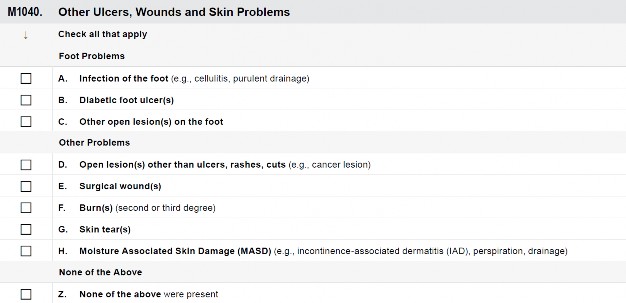
M1040: Other Ulcers, Wounds and Skin Problems
Skin wounds and lesions affect quality of life for residents because they may limit activity, may be painful, and may require time-consuming treatments and dressing changes.
Many of these ulcers, wounds and skin problems can worsen or increase risk for local and systemic infections.
This list represents only a subset of skin conditions or changes that nursing homes will assess and evaluate in residents.
The presence of wounds and skin changes should be accounted for in the interdisciplinary care plan.
This information identifies residents at risk for further complications or skin injury.
M1040: Other Ulcers, Wounds and Skin Problems (cont.)
DEFINITIONS
DIABETIC FOOT ULCERS
Ulcers caused by the neuropathic and small blood vessel complications of diabetes. Diabetic foot ulcers typically occur over the plantar (bottom) surface of the foot on load bearing areas such as the ball of the foot. Ulcers are usually deep, with necrotic tissue, moderate amounts of exudate, and callused wound edges. The wounds are very regular in shape and the wound edges are even with a punched-out appearance.
These wounds are typically not painful.
SURGICAL WOUNDS
Any healing and non-healing, open or closed surgical incisions, skin grafts or drainage sites.
OPEN LESION(S) OTHER THAN ULCERS, RASHES, CUTS
Most typically skin lesions that develop as a result of diseases and conditions such as syphilis and cancer.
BURNS (SECOND OR THIRD DEGREE)
Skin and tissue injury caused by heat or chemicals and may be in any stage of healing.
DEFINITIONS
DIABETIC FOOT ULCERS
Ulcers caused by the neuropathic and small blood vessel complications of diabetes. Diabetic foot ulcers typically occur over the plantar (bottom) surface of the foot on load bearing areas such as the ball of the foot. Ulcers are usually deep, with necrotic tissue, moderate amounts of exudate, and callused wound edges. The wounds are very regular in shape and the wound edges are even with a punched-out appearance.
These wounds are typically not painful.
SURGICAL WOUNDS
Any healing and non-healing, open or closed surgical incisions, skin grafts or drainage sites.
OPEN LESION(S) OTHER THAN ULCERS, RASHES, CUTS
Most typically skin lesions that develop as a result of diseases and conditions such as syphilis and cancer.
BURNS (SECOND OR THIRD DEGREE)
Skin and tissue injury caused by heat or chemicals and may be in any stage of healing.
Review the medical record, including skin care flow sheets or other skin tracking forms.
Speak with direct care staff and the treatment nurse to confirm conclusions from the medical record review.
Examine the resident and determine whether any ulcers, wounds, or skin problems are present.
Key areas for diabetic foot ulcers include the plantar (bottom) surface of the foot, especially the metatarsal heads (the ball of the foot).
Check all that apply in the last 7 days. If there is no evidence of such problems in the last 7 days, check none of the above.
Pressure ulcers/injuries coded in items M0210 through M0300 should not be coded here.
Check all that apply in the last 7 days. If there is no evidence of such problems in the last 7 days, check none of the above.
Pressure ulcers/injuries coded in items M0210 through M0300 should not be coded here.
M1040A, Infection of the foot (e.g., cellulitis, purulent drainage)
M1040B, Diabetic foot ulcer(s)
M1040C, Other open lesion(s) on the foot (e.g., cuts, fissures)
M1040D, Open lesion(s) other than ulcers, rashes, cuts (e.g., bullous pemphigoid)
M1040E, Surgical wound(s)
M1040F, Burn(s)(second or third degree)
M1040G, Skin tear(s)
M1040H, Moisture Associated Skin Damage (MASD) (e.g., incontinence-associated dermatitis (IAD), perspiration, drainage)
M1040Z, None of the above were present
M1040: Other Ulcers, Wounds and Skin Problems (cont.)
Diabetic neuropathy affects the lower extremities of individuals with diabetes. Individuals with diabetic neuropathy can have decreased awareness of pain in their feet. This means they are at high risk for foot injury, such as burns from hot water or heating pads, cuts or scrapes from stepping on foreign objects, and blisters from inappropriate or tight-fitting shoes. Because of decreased circulation and sensation, the resident may not be aware of the wound.
Neuropathy can also cause changes in the structure of the bones and tissue in the foot. This means the individual with diabetes experiences pressure on the foot in areas not meant to bear pressure. Neuropathy can also cause changes in normal sweating, which means the individual with diabetes can have dry, cracked skin on their other foot.
Do not include pressure ulcers/injuries that occur on residents with diabetes mellitus here. For example, an ulcer caused by pressure on the heel of a diabetic resident is a pressure ulcer and not a diabetic foot ulcer.
Open lesions that develop as part of a disease or condition and are not coded elsewhere on the MDS, such as wounds, boils, cysts, and vesicles, should be coded in this item.
Do not code rashes, abrasions, or cuts/lacerations here. Although not recorded on the MDS assessment, these skin conditions should be considered in the plan of care.
Do not code pressure ulcers/injuries, venous or arterial ulcers, diabetic foot ulcers, or skin tears here. These conditions are coded in other items on the MDS.
This category does not include healed surgical sites and healed stomas or lacerations that require suturing or butterfly closure as surgical wounds. PICC sites, central line sites, and peripheral IV sites are not coded as surgical wounds.
Surgical debridement of a pressure ulcer does not create a surgical wound. Surgical debridement is used to remove necrotic or infected tissue from the pressure ulcer in order to facilitate healing. A pressure ulcer that has been surgically debrided should continue to be coded as a pressure ulcer.
Code pressure ulcers that require surgical intervention for closure with graft and/or flap procedures in this item (e.g., excision of pressure ulcer with myocutaneous flap). Once a pressure ulcer is excised and a graft and/or flap is applied, it is no longer considered a pressure ulcer, but a surgical wound.
M1040: Other Ulcers, Wounds and Skin Problems (cont.)
Do not include first degree burns (changes in skin color only).
Skin tears are a result of shearing, friction or trauma to the skin that causes a separation of the skin layers. They can be partial or full thickness. Code all skin tears in this item, even if already coded in Item J1900B.
Do not code cuts/lacerations or abrasions here. Although not recorded on the MDS, these skin conditions should be considered in the plan of care.
DEFINITION
MOISTURE ASSOCIATED SKIN DAMAGE
Is superficial skin damage caused by sustained exposure to moisture such as incontinence, wound exudate, or perspiration.
DEFINITION
MOISTURE ASSOCIATED SKIN DAMAGE
Is superficial skin damage caused by sustained exposure to moisture such as incontinence, wound exudate, or perspiration.
MASD is also referred to as maceration and includes incontinence-associated dermatitis, intertriginous dermatitis, periwound moisture-associated dermatitis, and peristomal moisture-associated dermatitis.
Moisture exposure and MASD are risk factors for pressure ulcer/injury development. Provision of optimal skin care and early identification and treatment of minor cases of MASD can help avoid progression and skin breakdown.
MASD without skin erosion is characterized by red/bright red color (hyperpigmentation), and the
surrounding skin may be white (hypopigmentation). The skin damage is usually blanchable and diffuse and has irregular edges. Inflammation of the skin may also be present.
MASD with skin erosion has superficial/partial thickness skin loss and may have hyper- or hypopigmentation; the tissue is blanchable and diffuse and has irregular edges. Inflammation of the skin may also be present. Necrosis is not found in MASD.
If pressure and moisture are both present, code the skin damage as a pressure ulcer/injury in M0300.
If there is tissue damage extending into the subcutaneous tissue or deeper and/or necrosis is present, code the skin damage as a pressure ulcer in M0300.
M1040: Other Ulcers, Wounds and Skin Problems (cont.)
A resident with diabetes mellitus presents with an ulcer on the heel that is due to pressure.
Coding: This ulcer is not checked at M1040B. This ulcer should be coded where appropriate under the Pressure Ulcers items (M0210–M0300).
Rationale: Persons with diabetes can still develop pressure ulcers.
A resident is readmitted from the hospital after myocutaneous flap surgery to excise and close their sacral pressure ulcer.
Coding: Check M1040E, Surgical Wound.
Rationale: A surgical flap procedure was used to close the resident’s pressure ulcer. The pressure ulcer is now considered a surgical wound.
Resident J was reaching over to get a magazine off of their bedside table and sustained a skin tear on their wrist from the edge of the table when they pulled the magazine back towards them.
Coding: Check M1040G, Skin Tear(s).
Rationale: The resident sustained a skin tear while reaching for a magazine.
Resident S who is incontinent, is noted to have a large, red and excoriated area on their buttocks and interior thighs with serous exudate which is starting to cause skin glistening.
Coding: Check M1040H, Moisture Associated Skin Damage (MASD). Rationale: Resident S skin assessment reveals characteristics of incontinence- associated dermatitis.
Resident F complained of discomfort of their right great toe and when their stocking and shoe was removed, it was noted that their toe was red, inflamed and had pus draining from the edge of their nail bed. The podiatrist determined that Resident F has an infected ingrown toenail.
Coding: Check M1040A, Infection of the foot.
Rationale: Resident F has an infected right great toe due to an ingrown toenail.
Resident G has bullous pemphigoid and requires the application of sterile dressings to the open and weeping blistered areas.
Coding: Check M1040D, Open lesion other than ulcers, rashes, cuts.
Rationale: Resident G has open bullous pemphigoid blisters.
Resident A was just admitted to the nursing home from the hospital burn unit after sustaining second and third degree burns in a house fire. They are here for continued treatment of their burns and for rehabilitative therapy.
Coding: Check M1040F, Burns (second or third degree).
Rationale: Resident A has second and third degree burns, therefore, burns (second or third degree) should be checked.
M1200: Skin and Ulcer/Injury Treatments
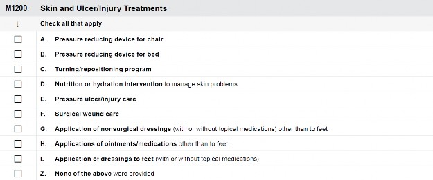
DEFINITION
PRESSURE REDUCING DEVICE(S)
Equipment that aims to relieve pressure away from areas of high risk. May include foam, air, water gel, or other cushioning placed on a chair, wheelchair, or bed.
Include pressure relieving, pressure reducing, and pressure redistributing devices. Devices are available for use with beds and seating.
DEFINITION
PRESSURE REDUCING DEVICE(S)
Equipment that aims to relieve pressure away from areas of high risk. May include foam, air, water gel, or other cushioning placed on a chair, wheelchair, or bed.
Include pressure relieving, pressure reducing, and pressure redistributing devices. Devices are available for use with beds and seating.
Appropriate prevention and treatment of skin changes and ulcers reduce complications and promote healing.
These general skin treatments include basic pressure ulcer/injury prevention and skin health interventions that are a part of providing quality care and consistent with good clinical practice for those with skin health problems.
These general treatments should guide more individualized and specific interventions in the care plan.
If skin changes are not improving or are worsening, this information may be helpful in determining more appropriate care.
Review the medical record, including treatment records and health care provider orders for documented skin treatments during the past 7 days. Some skin treatments may be part of routine standard care for residents, so check the nursing facility’s policies and procedures and indicate here if administered during the look-back period.
Speak with direct care staff and the treatment nurse to confirm conclusions from the medical record review.
Some skin treatments can be determined by observation. For example, observation of the resident’s wheelchair and bed will reveal if the resident is using pressure-reducing devices for the bed or wheelchair.
M1200: Skin and Ulcer/Injury Treatments (cont.)
Check all that apply in the last 7 days. Check Z, None of the above were provided, if none applied in the past 7 days.
Check all that apply in the last 7 days. Check Z, None of the above were provided, if none applied in the past 7 days.
M1200A, Pressure reducing device for chair
M1200B, Pressure reducing device for bed
M1200C, Turning/repositioning program
M1200D, Nutrition or hydration intervention to manage skin problems
M1200E, Pressure ulcer/injury care
M1200F, Surgical wound care
M1200G, Application of non-surgical dressings (with or without topical medications) other than to feet. Non- surgical dressings do not include Band-Aids.
M1200H, Application of ointments/medications other than to feet
M1200I, Application of dressings to feet (with or without topical medications)
M1200Z, None of the above were provided
M1200: Skin and Ulcer/Injury Treatments (cont.)
DEFINITIONS
TURNING/ REPOSITIONING PROGRAM
Includes a consistent program for changing the resident’s position and realigning the body. “Program” is defined as a specific approach that is organized, planned, documented, monitored, and evaluated based on an assessment of the resident’s needs.
NUTRITION OR HYDRATION INTERVENTION TO MANAGE SKIN PROBLEMS
Dietary measures received by the resident for the purpose of preventing or treating specific skin conditions, e.g., wheat-free diet to prevent allergic dermatitis, high calorie diet with added supplementation to prevent skin breakdown, high-protein supplementation for wound healing.
DEFINITIONS
TURNING/ REPOSITIONING PROGRAM
Includes a consistent program for changing the resident’s position and realigning the body. “Program” is defined as a specific approach that is organized, planned, documented, monitored, and evaluated based on an assessment of the resident’s needs.
NUTRITION OR HYDRATION INTERVENTION TO MANAGE SKIN PROBLEMS
Dietary measures received by the resident for the purpose of preventing or treating specific skin conditions, e.g., wheat-free diet to prevent allergic dermatitis, high calorie diet with added supplementation to prevent skin breakdown, high-protein supplementation for wound healing.
Pressure reducing devices redistribute pressure so that there is some relief on or near the area of the ulcer/injury. The appropriate pressure reducing device should be selected based on the individualized needs of the resident.
Do not include egg crate cushions of any type in this category.
Do not include doughnut or ring devices in chairs.
M1200C Turning/Repositioning Program
The turning/repositioning program is specific as to the approaches for changing the resident’s position and realigning the body. The program should specify the intervention (e.g., reposition on side, pillows between knees) and frequency (e.g., every 2 hours).
Progress notes, assessments, and other documentation (as dictated by facility policy) should support that the turning/repositioning program is monitored and reassessed to determine the effectiveness of the intervention.
M1200D Nutrition or Hydration Intervention to Manage Skin Problems
The determination as to whether or not one should receive nutritional or hydration interventions for skin problems should be based on an individualized nutritional assessment. The interdisciplinary team should review the resident’s diet and determine if the resident is taking in sufficient amounts of nutrients and
fluids or are already taking supplements that are fortified with the US Recommended Daily Intake (US RDI) of nutrients.
M1200: Skin and Ulcer/Injury Treatments (cont.)
Additional supplementation above the US RDI has not been proven to provide any further benefits for management of skin problems including pressure ulcers/injuries. Vitamin and mineral supplementation should only be employed as an intervention for managing skin problems, including pressure ulcers/injuries, when nutritional deficiencies are confirmed or suspected through a thorough nutritional assessment. If it is determined that nutritional supplementation, that is, adding additional protein, calories, or nutrients is warranted, the facility should document the nutrition or hydration factors that are influencing skin problems and/or wound healing and tailor nutritional supplementation to the individual’s intake, degree of under-nutrition, and relative impact of nutrition as a factor overall; and obtain dietary consultation as needed.
It is important to remember that additional supplementation is not automatically required for pressure ulcer/injury management. Any interventions should be specifically tailored to the resident’s needs, condition, and prognosis.
Pressure ulcer care includes any intervention for treating pressure ulcers coded in Current Number of Unhealed Pressure Ulcers/Injuries at Each Stage (M0300A–G). Examples may include the use of topical dressings; enzymatic, mechanical or surgical debridement; wound irrigations; negative pressure wound therapy (NPWT); and/or hydrotherapy.
Does not include post-operative care following eye or oral surgery.
Surgical debridement of a pressure ulcer does not create a surgical wound. Surgical debridement is used to remove necrotic or infected tissue from the pressure ulcer in order to facilitate healing, and thus, any wound care associated with pressure ulcer debridement would be coded in M1200E, Pressure Ulcer Care. The only time a surgical wound would be created is if the pressure ulcer itself was excised and a flap and/or graft used to close the pressure ulcer.
Surgical wound care may include any intervention for treating or protecting any type of surgical wound. Examples may include topical cleansing, wound irrigation, application of antimicrobial ointments, application of dressings of any type, suture/staple removal, and warm soaks or heat application.
Surgical wound care for pressure ulcers that require surgical intervention for closure (e.g., excision of pressure ulcer with flap and/or graft coverage) can be coded in this item, as once a pressure ulcer is excised and flap and/or graft applied, it is no longer considered a pressure ulcer, but a surgical wound.
M1200: Skin and Ulcer/Injury Treatments (cont.)
Do not code application of non-surgical dressings for pressure ulcers/injuries other than to feet in this item; use M1200E, Pressure ulcer/injury care.
Dressings do not have to be applied daily in order to be coded on the MDS assessment. If any dressing meeting the MDS definitions was applied even once during the 7-day look- back period, the assessor should check that MDS item.
This category may include, but is not limited to, dry gauze dressings, dressings moistened with saline or other solutions, transparent dressings, hydrogel dressings, and dressings with hydrocolloid or hydroactive particles used to treat a skin condition, compression bandages, etc. Non-surgical dressings do not include adhesive bandages (e.g., BAND- AID® bandages, wound closure strips).
Do not code application of ointments/medications (e.g., chemical or enzymatic debridement) for pressure ulcers here; use M1200E, Pressure ulcer/injury care.
This category may include ointments or medications used to treat a skin condition (e.g., cortisone, antifungal preparations, chemotherapeutic agents).
Ointments/medications may include topical creams, powders, and liquid sealants used to treat or prevent skin conditions.
This category does not include ointments used to treat non-skin conditions (e.g., nitropaste for chest pain, testosterone cream).
Includes interventions to treat any foot wound or ulcer other than a pressure ulcer/injury.
Do not code application of dressings to pressure ulcers/injuries on the foot; use M1200E, Pressure ulcer/injury care.
Do not code application of dressings to the ankle. The ankle is not considered part of the foot.
M1200: Skin and Ulcer/Injury Treatments (cont.)
A resident is admitted with a Stage 3 pressure ulcer on the sacrum. Care during the last 7 days has included one debridement by the wound care consultant, application of daily dressings with enzymatic ointment for continued debridement, nutritional supplementation, and use of a pressure reducing pad on the resident’s wheelchair. The medical record documents delivery of care and notes that the resident is on a two-hour turning/repositioning program that is organized, planned, documented, monitored, and evaluated based on an individualized assessment of their needs. The physician documents, after reviewing the resident’s nutritional intake, healing progress of the resident’s pressure ulcer, dietitian’s nutritional assessment, and laboratory results, that the resident has protein-calorie malnutrition. In order to support proper wound healing, the physician orders an oral supplement that provides all recommended daily allowances for protein, calories, nutrients, and micronutrients. All mattresses in the nursing home are pressure reducing mattresses.
Coding: Check items M1200A, M1200B, M1200C, M1200D, and M1200E. Rationale: Interventions include pressure reducing pad on the wheelchair (M1200A) and pressure reducing mattress on the bed (M1200B), turning and repositioning program (M1200C), nutritional supplementation (M1200D), enzymatic debridement and application of dressings (M1200E).
A resident has a venous ulcer on the right leg. During the last 7 days the resident has had a three-layer compression-bandaging system applied once (orders are to reapply the compression bandages every 5 days). The resident also has a pressure reducing mattress and pad for the wheelchair.
Coding: Check items M1200A, M1200B, and M1200G.
Rationale: Treatments include pressure reducing mattress (M1200B) and pad (M1200A) in the wheelchair and application of the compression-bandaging system (M1200G).
Resident S has a diagnosis of right-sided hemiplegia from a previous stroke. As part of their assessment, it was noted that while in bed Resident S is able to tolerate pressure on each side for approximately 3 hours before showing signs of the effects of pressure on their skin. Staff assist them to turn every 3 hours while in bed. When they are in their wheelchair, it is difficult for them to offload the pressure to their buttocks. Their assessment indicates that their skin cannot tolerate pressure for more than 1 hour without showing signs of the effect of the pressure when they are sitting, and therefore,
Resident S is assisted hourly by staff to stand for at least 1 full minute to relieve pressure. Staff document all of these interventions in the medical record and note the resident’s response to the interventions.
Coding: Check M1200C.
Rationale: Treatments meet the criteria for a turning/repositioning program (i.e., it is organized, planned, documented, monitored, and evaluated), that is based on an assessment of the resident’s unique needs.
M1200: Skin and Ulcer/Injury Treatments (cont.)
Resident J has a diagnosis of Advanced Alzheimer’s and is totally dependent on staff for all of their care. Their care plan states that they are to be turned and repositioned, per facility policy, every 2 hours.
Coding: Do not check item M1200C.
Rationale: Treatments provided do not meet the criteria for a turning/repositioning program. There is no notation in the medical record about an assessed need for turning/repositioning, nor is there a specific approach or plan related to positioning and realigning of the body. There is no reassessment of the resident’s response to turning and repositioning. There are not any skin or ulcer treatments being provided.
Scenarios for Pressure Ulcer Coding
Resident P was admitted to the nursing home on 10/23/2019 for a Medicare stay. In completing the PPS 5-day assessment (ARD of 10/28/2019), it was noted that the resident had a head-to-toe skin assessment and their skin was intact, but upon assessment using the Braden scale, was found to be at risk for skin breakdown. The resident was noted to have a Stage 2 pressure ulcer that was identified on their coccyx on 11/1/2019. This Stage 2 pressure ulcer was noted to have pink tissue with some epithelialization present in the wound bed. Dimensions of the ulcer were length 01.1 cm, width 00.5 cm, and no measurable depth. Resident P does not have any arterial or venous ulcers, wounds, or skin problems. They are receiving ulcer care with application of a dressing applied to the coccygeal ulcer. Resident P also has pressure reducing devices on both their bed and chair and has been placed on a 1½ hour turning and repositioning schedule per tissue tolerance. In order to stay closer to their family, Resident P was discharged to another nursing home on 11/5/2019. This was a planned discharge (A0310G = 2), and their OBRA Discharge assessment was coded at A0310F as 10, Discharge assessment – return not anticipated.
Day PPS:
Coding:
Rationale: The resident has a pressure ulcer. On the 5-day PPS assessment, the resident’s skin was noted to be intact; however, on the Discharge assessment, it was noted that the resident had a new Stage 2 pressure ulcer. Since the resident has had both a 5-day PPS and Discharge assessment completed, the Discharge assessment would be coded 0 at A0310E. This is because the Discharge assessment is not the first assessment since the most recent admission/entry or reentry.